Ultrasound During Pregnancy: Are They Dangerous?
All pregnant women long to know how their children are doing and ultrasounds are necessary to find out. They give us the opportunity to see the condition of the fetus and detect any abnormalities or problems. However, there are some who claim that ultrasound examinations during pregnancy are dangerous for the unborn baby.
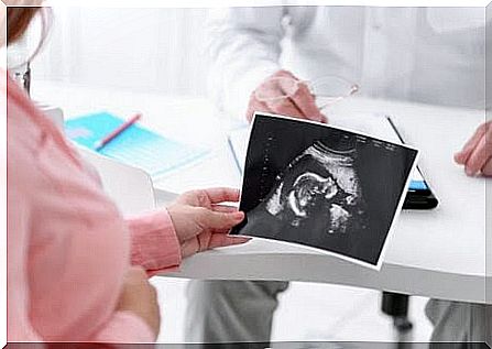
If this is the case for you, it is a good idea to talk to your doctor and ask him or her to explain the real implications of ultrasound.
What are ultrasounds and what are they used for?
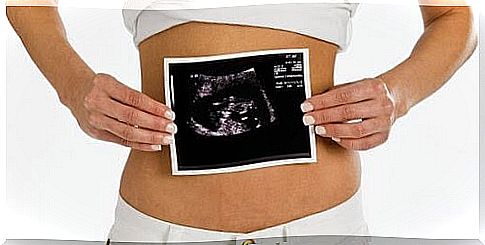
Ultrasound consists of images created by waves transmitted from the abdomen or through the vagina. These waves bounce from the uterus and placenta to produce the image.
An examination with ultrasound can be between 15 and 20 minutes. These are used to confirm that the baby is alive and also measure the amount of amniotic fluid that is present. They are also useful for detecting twin pregnancies and checking for any umbilical cord abnormalities.
Ultrasound images also provide information regarding the growth and weight of the fetus, the position of the placenta and serious complications, such as placenta praevia . But the most important thing is that they help to detect any problems that may lead to premature birth.
Why do some people say that ultrasound examinations during pregnancy are dangerous?
There is a myth that it is dangerous to do ultrasound examinations during pregnancy. Those who say this claim that the ultrasound waves can increase the temperature in the tissues.
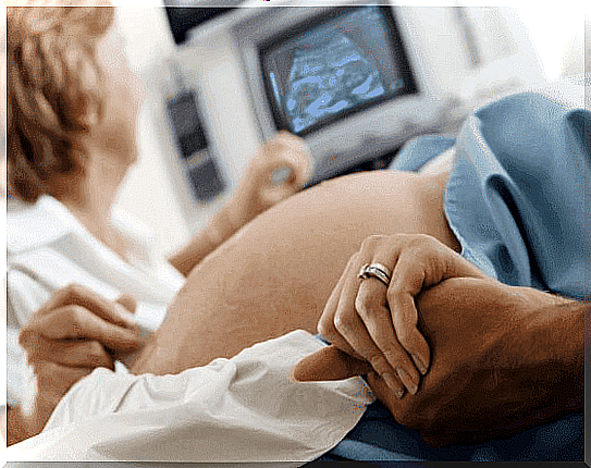
But the technology is completely harmless and very simple. It does not pose a danger at all to the fetus or to the mother, given that it does not use ionizing radiation, such as X-rays. In fact, ultrasound images do not cause any changes or sudden movements in the fetus at all.
However, one should not take it too far but instead only turn to ultrasound as it is recommended by a doctor.
How many ultrasound examinations do doctors recommend?
It is perfectly normal to have an ultrasound examination during pregnancy and there is no risk at all for pregnant women or their unborn children.
In larger parts of the world, doctors normally recommend three ultrasounds – one during each trimester. The first is usually between weeks 11 and 14, the second between weeks 18 and 22 and the last between weeks 32 and 36. In Sweden, it differs between different county councils and municipalities, but you are always called in for at least one ultrasound around week 20.
In high-risk pregnancies, where there is a higher risk of miscarriage, hypertension, diabetes or other complications, doctors may recommend additional ultrasound examinations. In some cases, doctors may even recommend ultrasound up to every two weeks.
Should the woman have a full bladder at the examination?
The answer depends on whether the ultrasound is via the abdomen during the first weeks of pregnancy. In this case, the doctor usually recommends that you drink several glasses of water before to get better pictures.
However, in transvaginal ultrasound examinations, it is not necessary to drink water. After 12 weeks of pregnancy, it is not necessary to drink water for ultrasound through the abdomen either because there is enough amniotic fluid to get nice pictures.
Types of ultrasound during pregnancy
There are different types of ultrasounds that allow parents and doctors to see the growing fetus.
2D ultrasound
As mentioned above, you usually take three ultrasounds during a pregnancy:
- Week 11-14. The first ultrasound provides information about the number of fetuses and how far along the pregnancy is. It also detects possible early abnormalities, as well as the presence of fibroids and cysts in the mother.
- Week 18-20. In addition to revealing the baby’s position and the size of the fetus, ultrasound at this time can verify the baby’s anatomy and hence gender. Doctors can observe the baby’s head, spine, heart and extremities.
- Week 32-36. During the third trimester, an ultrasound again shows the baby’s position and also measures the amount of amniotic fluid that is present. In addition, it can detect abnormalities that appear later, such as kidney obstructions and water skull.
3D ultrasound
With these pictures you can see your child’s face, body and internal structures more accurately. They also help confirm any abnormalities previously detected during a 2D ultrasound.
4D ultrasound
This advanced technology detects skin problems and deformities, such as cleft lip. It also shows the child’s movements in real time and shows the child’s face in greater detail. This means that parents can get a clear idea of their child’s appearance.
In summary, you can be sure that ultrasound during pregnancy is harmless. If you are pregnant, do not be afraid to go for all the ultrasound examinations that your doctor recommends for you.